
How Diabetes Secretly Damages Your Eyes: Early Warning Signs of Diabetic Retinopathy You Shouldn't Ignore
Diabetic retinopathy is the leading cause of preventable blindness in adults. Learn how diabetes silently affects retinal micro-vessels, why 20/20 vision can hide dangerous bleeding, and how early laser or anti-VEGF therapy saves sight.

- You can have severe diabetic retinal capillary damage even while your vision remains completely sharp and 20/20.
- High blood sugar weakens tiny retinal blood vessels, causing microaneurysms that leak protein, fluid, and blood into the macula.
- Diabetic Macular Edema (DME) is the most common cause of vision loss in working-age diabetics and can be reversed if treated promptly with Anti-VEGF injections.
- Every person diagnosed with Type 2 diabetes must undergo a dilated retina examination and OCT scan at least once every 12 months.
- Strict control of HbA1c (below 7%), blood pressure (<130/80 mmHg), and serum lipids drastically reduces your risk of retinal hemorrhage.
1. The Silent Menace Behind 'Normal' Vision
In our retina clinic at Ashvini Netralaya, we frequently meet patients who say: 'Doctor, I have had diabetes for 12 years, but my eyesight was perfect until last week when sudden black spots appeared. How could my eyes deteriorate so fast?'
The sobering truth is that diabetic retinopathy does not develop overnight. It progresses silently over years. The retina—the light-sensitive neurosensory wallpaper lining the back of your eye—is packed with microscopic capillaries.
Chronic exposure to elevated blood glucose damages the delicate walls of these capillaries (pericyte loss). They balloon out into microaneurysms, leak plasma and lipids, and eventually starve parts of the retina of oxygen.
Because your peripheral retina often suffers damage first while the central macula remains untouched in early stages, you may notice zero blurriness while dangerous changes are underway.
2. The Stages of Diabetic Retinopathy
Ophthalmologists classify diabetic eye disease into two broad progressive stages:
**1. Non-Proliferative Diabetic Retinopathy (NPDR)**: In this initial stage, retinal blood vessels leak fluid and tiny dots of blood (dot-and-blot hemorrhages). It ranges from mild to moderate and severe.
**2. Proliferative Diabetic Retinopathy (PDR)**: When capillary blockages starve large areas of the retina of oxygen (ischemia), the eye panics and releases abnormal growth factors (VEGF). This stimulates the growth of fragile, abnormal new blood vessels (neovascularization). These abnormal vessels bleed easily into the vitreous cavity (vitreous hemorrhage) and can tug on the retina, causing a tractional retinal detachment.
| Stage | Symptoms | Clinical Findings | Standard Management |
|---|---|---|---|
| Mild NPDR | None | Isolated microaneurysms | HbA1c optimization, annual dilated checkup |
| Moderate/Severe NPDR | Mild fluctuations in focus | Widespread hemorrhages, cotton wool spots | Dilated exam every 4-6 months, OCT scan |
| Diabetic Macular Edema (DME) | Central blur, wavy straight lines | Fluid swelling in central retina | Intravitreal Anti-VEGF injections (Lucentis/Eylea/Avastin) |
| Proliferative Retinopathy (PDR) | Sudden floaters, cobwebs, red haze | Fragile new vessels, vitreous bleed | Pan-Retinal Green Laser (PRP) + Anti-VEGF or Vitrectomy |
3. Diabetic Macular Edema (DME): Why Things Look Wavy
The macula is the center of your retina responsible for sharp, reading vision. When leaky diabetic vessels cause fluid swelling within the macula, your vision becomes blurred, colors look washed out, and straight lines (such as door frames or graph paper) appear wavy or distorted.
At Ashvini Netralaya, we diagnose DME using the gold-standard Carl Zeiss Cirrus HD-OCT (Optical Coherence Tomography). In less than 3 seconds, this non-invasive laser scan generates microscopic cross-sectional images of your retinal layers, measuring fluid accumulation down to the single micron level.
The Home Amsler Grid Test
Diabetic patients should check each eye separately once a week using an Amsler grid (or a bathroom tile pattern). If straight grid lines appear bent, wavy, or missing, contact your retina specialist immediately.
4. Modern Treatments: Injections, Lasers, and Vitrectomy
A diagnosis of diabetic retinopathy no longer means inevitable blindness. Over the past decade, vitreoretinal medicine has undergone a revolutionary transformation:
**1. Anti-VEGF Injections**: Painless, microscopic injections given inside the vitreous cavity using numbing eye drops. Medications such as Ranibizumab, Aflibercept, and Bevacizumab block abnormal leakage, rapidly drying up macular swelling and restoring clarity.
**2. 532nm Green Laser (Pan-Retinal Photocoagulation)**: We use targeted laser energy to seal off ischemic peripheral retinal zones. This stops the release of VEGF and permanently shrinks dangerous new vessels, preventing catastrophic hemorrhages.
**3. 25-Gauge/27-Gauge Micro-Incision Vitrectomy (MIVS)**: For patients with non-clearing vitreous bleeding or retinal detachment, our surgical team performs sutureless vitrectomy, delicately clearing blood and relieving retinal traction under high-magnification surgical operating microscopes.
5. The Golden Rule of Diabetic Eye Protection
Treating the eye without managing your systemic health is like baling water out of a boat while the leak remains open. Clinical trials prove that maintaining an HbA1c below 7.0%, controlling systolic blood pressure below 130 mmHg, and keeping kidney parameters healthy reduces retinopathy progression by over 70%.
Do not wait for your vision to fail. If you have diabetes, schedule your comprehensive dilated retina examination today.
Frequently Asked Patient Questions
Does an eye injection for diabetes hurt?
How often does a diabetic need an eye checkup?
Can laser treatment restore lost vision?
Ashvini Netralaya • Superspeciality Eye Hospital
Need a Specialist Eye Consultation or Second Opinion?
Ashvini Netralaya is Tripura's 1st and only NABH-accredited superspeciality eye hospital, offering state-of-the-art diagnostic and surgical eye care in central Agartala.
Related Clinical Treatments at Ashvini Netralaya
Retina Surgeries & Diabetic Eye Care
Advanced 25G/27G sutureless vitrectomy, Anti-VEGF pharmacotherapy, and green laser photocoagulation.
Lasers & High-Precision Diagnostics
Zeiss Cirrus HD-OCT and digital fundus photography for early microvascular leakage detection.
Recommended Eye Health Guides
View all articles →
Choosing the Right Lens for Cataract Surgery: Monofocal, Toric, or Multifocal? A Surgeon's Guide
Confused between Monofocal, Toric, Multifocal, and EDOF intraocular lenses (IOLs)? Here is an honest, clinical breakdown to help you pick the perfect lens for your lifestyle, budget, and vision goals.
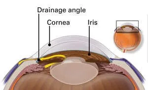
Glaucoma: The Silent Thief of Sight — Why Normal Vision Doesn't Always Mean Healthy Eyes
Glaucoma damages your optic nerve without causing pain, red eyes, or noticeable blurriness until 80% of fibers are permanently destroyed. Learn the risk factors, the importance of eye pressure screening, and modern pressure-lowering therapies.